
 Français
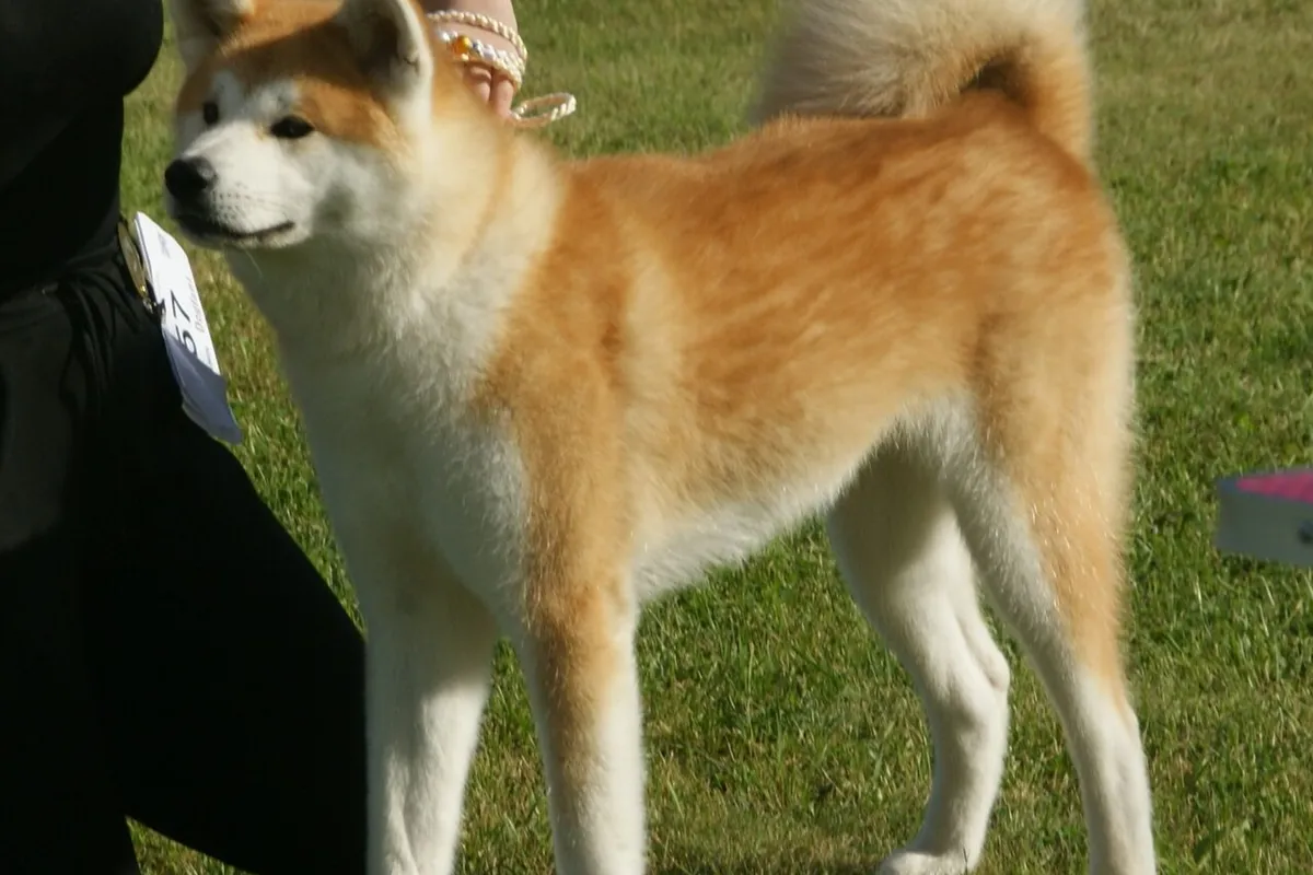
At a glance
| Trait | Detail |
|---|---|
| Origin | Japan [1] |
| Breed group | FCI group 5, section 5 (Asian Spitz and related breeds) [1]. The American Akita is a separate FCI breed in the same section [2] |
| Weight | Median adult weight of dogs recorded as Akitas in UK practices 88 lb (40.1 kg): females 83 lb (37.6 kg), males 94 lb (42.7 kg) [3] |
| Height | Males 26.5 in (67 cm), females 24 in (61 cm), with a tolerance of 3 cm (about 1 in) either way [1] |
| Life expectancy | Median age at death 11.3 years in UK veterinary records [3]; median 11.4 years in a large UK study that grouped Japanese and American Akitas [4] |
| Coat | Harsh, straight outer coat over a soft, dense undercoat; red fawn, sesame, brindle or white, with whitish “urajiro” markings except in white dogs [1] |
Origin
From 1603, medium-sized bear-hunting dogs called Akita Matagis were used as fighting dogs in the Akita region of Japan. From 1868 they were crossed with Tosas and Mastiffs, which increased their size. Dog fighting was banned in 1908, the breed was kept as a large Japanese breed, and nine dogs were designated “Natural Monuments” in 1931. After the Second World War, breeders crossed back to Matagi Akitas to restore the original type [1].
Akitas of the Dewa line, which showed Mastiff and German Shepherd influence, were taken to the United States by members of the armed forces after the war. They became the American Akita, now recognized by the FCI as a separate breed [2]. This page is about the Japanese Akita; some studies below do not separate the two.
Common health problems
- Hypothyroidism: 0.76% of Japanese Akita Inus in UK practices were recorded with hypothyroidism, and the breed had 4.69 times the odds of crossbreeds [5]. See hypothyroidism in dogs.
- Gastric dilatation-volvulus (GDV, bloat): in UK emergency clinics, Akitas had 84.4 times the odds of crossbreeds of a GDV diagnosis, second only to the Great Dane [6]. See our Great Dane page.
- Uveodermatologic syndrome (VKH-like syndrome): an immune attack on pigmented cells of the eye and skin. In a study of 50 dogs with the disease, 37 were Akitas. At the first visit 57% were blind in both eyes and 36% had glaucoma, but 78% of the dogs that had or regained vision kept it until their last visit [8].
- Sebaceous adenitis: an immune skin disease that destroys the oil glands of the skin. In a study of 97 purebred Akitas, 23 had the disease on biopsy. Lesions began mainly on the back and ears, hair loss affected mostly the undercoat, and an autosomal recessive inheritance appeared possible [7].
What is special in the Akita’s blood work
Two features of the Akita’s blood can make a healthy dog’s results look abnormal: potassium-rich red cells and low platelet counts.
| Test | Akitas with high red-cell potassium | Akitas with normal red cells |
|---|---|---|
| Plasma potassium | 6.6 mmol/L [11] | 4.4 mmol/L [11] |
| Hematocrit (PCV) | 42.9% [11] | 46.7% [11] |
| Hemoglobin | 13.5 g/dL [11] | 15.9 g/dL [11] |
| MCV | 70.0 fL [11] | 63.4 fL [11] |
Potassium. Dogs usually have little potassium inside their red blood cells, but some Japanese and other Asian breeds, such as the Akita and the Shiba Inu, have a lot [10]. In the first report, a healthy Akita with a high potassium had normal ACTH stimulation test results, and the high value was traced to hemolysis of the sample. Six of 8 Akitas tested had high red-cell potassium, and plasma potassium rose the longer the plasma stayed in contact with the cells, from 4 hours of refrigeration [9]. Separating serum or plasma from the cells quickly avoids this artifact [10]. See potassium in dogs and cats.
Red cells. Akitas with high red-cell potassium had a lower hematocrit, hemoglobin and MCHC and a higher MCV than other Akitas [11], while Akitas as a group had a lower MCV than control dogs in an Australian study [12]. Read with ordinary dog intervals, a healthy Akita’s red cell values can look slightly abnormal. See anemia in dogs.
Platelets. In 18 healthy Akitas in Australia, 83% had a platelet count below 150 × 10⁹/L, against none of 11 control dogs, and none showed bleeding. Automated counts were lower than manual counts, and large platelets were seen in only 11% of Akitas. The authors advise confirming low counts manually to avoid a wrong diagnosis of immune thrombocytopenia and needless treatment [12]. See low platelets in dogs.
Related articles
En français : Akita inu.
Questions to ask your veterinarian
- Could this high potassium be an artifact of my Akita’s red cells?
- Was the sample hemolyzed, or left long before the serum was separated?
- Has the low platelet count been checked by a manual count or on a smear?
- Do my dog’s thyroid results point to hypothyroidism?
- What are the signs of bloat, and what should I do if I see them?
Get your Akita’s blood work explained.
AI DiagAnimals reads potassium, the red cell values, platelets and thyroid results together with your laboratory’s intervals, and flags the values that often differ in Akitas. The service opens soon: leave your email to be told first.
Frequently asked questions
How long do Akitas live?
In UK veterinary records, Akitas that died did so at a median age of 11.3 years [3]. A large UK study grouping Japanese and American Akitas found a median of 11.4 years [4].
Why is my Akita’s potassium high?
Many Akitas have potassium-rich red blood cells. If the sample is hemolyzed or the serum is not separated quickly, potassium leaks out and the result looks high in a healthy dog [9, 10].
Is a low platelet count normal in Akitas?
It is common: 83% of healthy Akitas in an Australian study had counts below 150 × 10⁹/L without bleeding, and analyzers tended to undercount [12].
Are Akitas prone to hypothyroidism?
Yes. In UK practices, Japanese Akita Inus had 4.69 times the odds of crossbreeds [5].
Sources
- FCI. Standard No. 255: Akita. 2001. fci.be/Nomenclature/Standards/255g05-en.pdf
- FCI. Standard No. 344: American Akita. 2026. fci.be/Nomenclature/Standards/344g05-en.pdf
- O’Neill DG et al. Giant dog breeds under primary veterinary care in the UK: demography, common disorders and mortality. Companion Anim Health Genet. 2026;13:4. doi.org/10.1186/s40575-026-00152-7
- McMillan KM et al. Longevity of companion dog breeds: those at risk from early death. Sci Rep. 2024;14:531 (supplementary tables S3 and S5). doi.org/10.1038/s41598-023-50458-w
- O’Neill DG et al. Frequency, breed predispositions and other demographic risk factors for diagnosis of hypothyroidism in dogs under primary veterinary care in the UK. Canine Med Genet. 2022;9:11. doi.org/10.1186/s40575-022-00123-8
- O’Neill DG et al. Gastric dilation-volvulus in dogs attending UK emergency-care veterinary practices: prevalence, risk factors and survival. J Small Anim Pract. 2017;58(11):629-638. doi.org/10.1111/jsap.12723
- Reichler IM et al. Sebaceous adenitis in the Akita: clinical observations, histopathology and heredity. Vet Dermatol. 2001;12(5):243-253. doi.org/10.1046/j.0959-4493.2001.00251.x
- Zarfoss MK et al. Clinical findings and outcomes for dogs with uveodermatologic syndrome. J Am Vet Med Assoc. 2018;252(10):1263-1271. doi.org/10.2460/javma.252.10.1263
- Degen M. Pseudohyperkalemia in Akitas. J Am Vet Med Assoc. 1987;190(5):541-543. pubmed.ncbi.nlm.nih.gov/3558091
- eClinPath (Cornell University). Potassium. eclinpath.com/chemistry/electrolytes/potassium
- Conrado FO et al. Clinicopathologic and electrocardiographic features of Akita dogs with high and low erythrocyte potassium phenotypes. Vet Clin Pathol. 2014;43(1):50-54. doi.org/10.1111/vcp.12112
- Bool JD et al. Asymptomatic thrombocytopenia in Akitas: a prospective cross-sectional study of platelet characteristics in an Eastern Australian population. Aust Vet J. 2026;104:600-604. doi.org/10.1111/avj.70111
Laboratory methods and reference intervals vary. This article explains; it does not diagnose. For a decision about your pet, use the original report and talk to your veterinarian. Our editorial policy.